



Modeling the Extracellular Matrix-Like Retinal Protein Pikachurin and its Binding to
α-Dystroglycan and GPCR (GPR179)
Authors: Mohit Belur, Khushi Raval, Riya Chelagiri, Rani Mehta, Emily Schmitt Lavin, Arthur Sikora
School: Nova Southeastern University, Fort Lauderdale, FL
PBD ID: 7ZCB (without GPCR), 8D1B (GPCR attached to Pikachurin), 9C3C (DystrophinGlycoprotein Complex)
We developed a 3D printed model to better understand the structure and function of the molecule, Pikachurin. Pikachurin is an extracellular matrix scaffold protein that bridges photoreceptor and ON-bipolar cell synapses by linking the presynaptic dystroglycan complex to the postsynaptic receptor GPR179, ensuring precise synaptic alignment essential for normal vision. Pikachurin was discovered in 2008 by a Japanese team at the Osaka Bioscience Institute through a microarray analysis of mouse retinas and was named after the Pokémon, Pikachu, because of its “lightning-fast” function in vision transmission. Although its role in dystroglycanopathy associated visual phenotypes is well recognized, there is no published structural model showing how pikachurin engages α-dystroglycan or how this may influence GPCR linked synaptic signaling. The objective of this study was to computationally model a full length Pikachurin based on available Protein Data Bank (PDB) structures with a focus on evaluating the Ca²⁺ -binding domain as a potential site for glycan-mediated interaction with dystroglycan. Structural modeling using Jmol, AlphaFold and literature-based analysis were used to predict domain organization, Ca²⁺-binding motifs, and predicted interaction sites between Pikachurin with the disaccharide-modified dystroglycan. The analysis showed that Pikachurin contains two Fibronectin Type III domains and a Ca²⁺-binding site that enables high-affinity recognition of glycosylated dystroglycan, supporting its role as a synaptic “bridge” protein. These findings suggest that proper glycosylation of dystroglycan is essential for stable Pikachurin binding and synaptic connectivity, and disruptions in this interaction may lead to impaired synaptic transmission and visual function. Our 3D printed model is based on an AlphaFold structure that was produced using the full nucleic acid sequence of Pikachurin, which combines the Fibronectin Type III (FN3 I, FN3 II), and Laminin G-like (LG1, LG2, and LG3) domains. The polysaccharide from PDB ID 7ZCB is 3D printed and bound to the Asn47 binding site as indicated from the literature. A dystroglycan polysaccharide is also printed and attached to the expected calcium binding site between the LG1 and LG2 domains. The residues (within the LG3 domain) that bind with a GPCR (GPR179) in the to the postsynaptic membrane are also highlighted. Through the use of this 3D molecular model, we are better able to understand and explain the story of how Pikachurin is embedded in the extracellular matrix and connected to the presynaptic neuron.

Primary Citation:
-
Patil DN, Pantalone S, Cao Y, Laboute T, Novick SJ, Singh S, Savino S, Faravelli S, Magnani F, Griffin PR, Singh AK, Forneris F, Martemyanov KA. Structure of the photoreceptor synaptic assembly of the extracellular matrix protein pikachurin with the orphan receptor GPR179. Sci Signal. 2023 Jul 25;16(795):eadd9539. doi: 10.1126/scisignal.add9539. Epub 2023 Jul 25. PMID: 37490546; PMCID: PMC10561654. https://pubmed.ncbi.nlm.nih.gov/37490546/
Modeling the Disruption of Binding of Three IgE Antibodies with Six Mutated Interaction Sites on the Arah2 Allergen
Authors: Hansi Parmar, Aashi Chhabra, Jyothi Vivekananda, Amaan Khan, Arthur Sikora, Emily Schmitt Lavin
School: Nova Southeastern University, Fort Lauderdale, FL
PBD ID: 8G4P, 8DB4
Peanut allergy is a life-threatening food allergy that affects millions of children and adults across the world. This issue presents serious public health concerns and heavy interest from the scientific community to find viable treatments that minimize immunological side effects. To better understand and explain the peanut allergy response, the literature was examined, and a 3D model was printed based on two protein database files. Arachis hypogaea 2 (Arah2) is the dominant peanut allergen protein. When Arah2 binds IgE antibodies, a strong allergic response is triggered. This reaction makes treating allergy patients with Oral Immunotherapy (OIT) difficult due to adverse effects from allergic reactions.
Existing PDBs 8G4P and 8DB4 from the literature reveal three monoclonal antibodies (mAbs) which bind the dominant IgE binding sites to elicit this inflammatory allergic reaction cascade. The structural epitopes of each of these antibodies interact with specific amino acid residues on the Arah2 allergen. In these PDBs, six mutations on the allergen, E46R, E89R, E97R, E114R, Q146A, and R147E, were targeted to generate a hexamutant Arah2 hypoallergen. These modifications demonstrated reduced IgE reactivity while maintaining the native Arah2 structure. The study concludes that allergic reactions can be reduced through rational amino acid substitutions that disrupt antibody binding. The study provides a basis for immunotherapies by using structurally similar but less reactive allergens.
Here, we prepared a 3D model of the hexamutant hypoallergen using AlphaFold 3 to predict the hexamutant structure, while ensuring that the overall conformation was conserved. Using Pymol and Jmol, a 3D printed molecular model was prepared with the 8G4P and 8DB4 PDB files in order to dock three antibodies to the hexamutant structure.
This 3D printed model highlights the binding of mAbs 13T1, 13T5, and 22S1 to three different sites and demonstrates why the hypoallergen mutations disrupt binding while conserving Arah2’s tertiary structure. Molecular interactions are compared, including hydrogen bonds and possible ionic interactions, with specific attention to key amino acid mutations in the binding sites. Through this work, we show that details of the molecular stories in the protein data bank can be better visualized and understood with the development and use of 3D printed models.

Primary Citation:
-
Min J, Keswani T, LaHood NA, et al. Design of an Ara h 2 hypoallergen from conformational epitopes. Clin Exp Allergy. 2024; 54: 46-55. doi:10.1111/cea.14433
Modeling OCT3-Mediated Buprenorphine Transport as a Mechanism for Salivary Accumulation and Oral Toxicity
Authors: Veda Pesala, Laasya Vemparala, Mehtab Singh, Sydni Gunpath, Emily Schmitt Lavin, Arthur Sikora
School: Nova Southeastern University, Fort Lauderdale, FL
PBD ID: 7ZH0
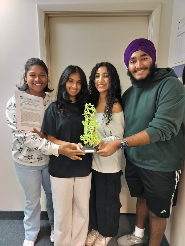
In this study we prepared a 3D printed model to better understand OCT3-mediated buprenorphine transport. Buprenorphine is a high affinity partial agonist at the mu-opioid receptor (μ-OR) and main component of Suboxone. It has a unique pharmacological profile that makes it effective for treating both pain and opioid use disorder (OUD) but has been linked to oral health issues such as dry mouth and tooth decay. The exceptionally tight binding of suboxone to μ-OR allows for sustained receptor occupancy and reduced opioid misuse potential by patients. Activation of μ-ORs can inhibit the release of acetylcholine. This neurotransmitter found in many cell types is essential for stimulating salivary secretion through muscarinic M3 receptors. When acetylcholine release is suppressed, calcium-dependent signaling in salivary gland cells decreases, leading to reduced water and ion secretion and, ultimately, dry mouth. Clinical studies have shown that buprenorphine and its metabolite, norbuprenorphine, reach concentrations up to 100 times higher in saliva than in plasma, suggesting the involvement of a cell specific active transport mechanism. Recent evidence suggests that the Organic Cation Transporter 3 (OCT3), a membrane transporter expressed in salivary gland epithelial cells, facilitates drug excretion into salivary glands. We investigated this interaction and its role in mediating buprenorphine movement into saliva. Using molecular docking and structural modeling of human OCT3 crystal structures (PDBIDs: 7ZH0, 7ZH6, 7ZHA), buprenorphine binding interactions within the central cavity of the transporter were analyzed. OCT3 has been found to transport many substrates that share structural similarities with buprenorphine, including metformin, morphine, corticosterone, and decynium-22. Docking studies revealed that buprenorphine exhibits a high binding affinity and specificity to OCT3. Although buprenorphine’s calculated affinity (−8.375 kcal/mol) is slightly lower than corticosterone (−10.640 kcal/mol) and decynium-22 (−9.749 kcal/mol), its unique interactions with PHE36, PHE250, ILE254, GLU451, and TYR454 support the hypothesis that this molecule has specific strong interactions with OCT3. These buprenorphine-specific residues are hypothesized to form strong hydrophobic interactions with the molecule, stabilizing polar interactions found with other substrates of the transporter. In contrast, metformin binds mainly to polar residues, while corticosterone, decynium-22, and morphine involve multiple shared aromatic residues, indicating broader but less selective binding. By understanding the mechanism of selective transport and salivary accumulation of buprenorphine, potential strategies to reduce its oral side effects while improving treatment outcomes for individuals with OUD can be identified.
Primary Citation:
-
Khanppnavar, B., Maier, J., Herborg, F., Gradisch, R., Lazzarin, E., Luethi, D., Yang, J., Qi, C., Holy, M., Jäntsch, K., Kudlacek, O., Schicker, K., Werge, T., Gether, U., Stockner, T., Korkhov, V.M., & Sitte, H.H. (2022). Structural basis of organic cation transporter-3 inhibition. Nature Communications, 13, 6714. https://doi.org/10.1038/s41467-022-34284-8
Modeling the Binding of Salinosporamide A (MZB) to the Human 20S Proteasome for Potential Cancer Treatment
Authors: Jayden J. Ross, Giezel Medina, Audrey Anand, Jayant Ari, Emily Schmitt, Arthur Sikora
School: Nova Southeastern University, Fort Lauderdale, FL
PBD ID: 9HMN
This project aims to model and observe the binding of the drug Marizomib (MZB) and compare its binding affinity to that of other similar drugs to the proteosome using the available PDB ID 9HMN and bioinformatics tools. MZB is an anti-cancer drug that was derived from the marine bacterium, Salinosporamide A. Its function is to inhibit the human 20S proteasome from breaking down misfolded and excess proteins, which eventually causes the cell to trigger apoptosis. Additionally, MZB has a unique ability to cross the blood-brain barrier and utilize three catalytically active amino acid sites. The existing PDB ID (9HMN) of a 20S proteasome with MZB bound was 3D printed. The catalytically inactive alpha subunits and one beta ring was removed to better model the three active beta sites. The catalytically active subunits, important binding residues, and the MZB drug were then color coded. To compare the binding of MZB to that of the similar drug Carfilzomib (CFZ), we used SwissDock to isolate the beta-5 subunit and dock Carfilzomib in the area where MZB typically binds. It was concluded that MZB and CFZ have similar binding regions of the beta-5 subunit, though at different residues. The MZB binding has greater affinity for the β1, β2, and β5 subunits, while the CFZ binds only to the β5-subunit site. It was determined that MZB specifically bound to the three catalytic “conserved triad” residues (Thr1, Asp17, and Lys33) on each of the three subunits. Further binding residues for MZB include Gly47, Thr21 and Ser130 for main and side chain interactions, and Ser129, Asp166, and Ser169 for structural integrity. Based on the locations where the drug binds, the MZB drug shows the highest affinities for the subunits and thus has the greatest applications for cancer treatments. The process of modeling the proteasome inhibitor MZB and the use of a 3D printed model helps illustrate its potential for cancer treatments (including glioblastoma and myeloma). MZB can bind to all three of the catalytically active subunits within the beta-ring of the proteasome, which is something that the other drugs we investigated cannot do effectively. Furthermore, MZB is stabilized within the active site by binding to the conserved triad and other residues for structural integrity.

Primary Citation:
-
Sülzen, H., Fajtova, P., O'Donoghue, A. J., Silhan, J., & Boura, E. (2025). Structural Insights into Salinosporamide a Mediated Inhibition of the Human 20S Proteasome. Molecules (Basel, Switzerland), 30(6), 1386. https://doi.org/10.3390/molecules30061386
Molecular Consequences of PCOS-Linked Aromatase Variants: Structural Modeling and Inhibitor Docking
Authors: Kiran S. Sajnani, Bharath Kumar Reddy Burri, Yuktha Milukuri, Preya Patel, Marqus Colón, Emily Schmitt Lavin Ph.D., Arthur Sikora Ph.D.
School: Nova Southeastern University, Fort Lauderdale, FL
PBD ID: 3EQM
Polycystic ovary syndrome (PCOS) affects approximately 10% of women of reproductive age and involves hormonal imbalances partly driven by disrupted estrogen synthesis. Aromatase (CYP19A1) catalyzes the conversion of testosterone and androstenedione into estrogen, making it essential for maintaining estrogen–androgen balance. The structural consequences of disease-associated aromatase mutations remain poorly understood. We investigated the Val161Asp substitution, a PCOS-linked variant in which a nonpolar valine is replaced by a negatively charged aspartate within a hydrophobic helical region of aromatase. Using the wild-type structure from PDB ID 3EQM, we generated a 3D-printed model highlighting the mutation and identified key catalytic features, including the heme prosthetic group, substrate-binding channel, and conserved monooxygenase motifs. We then produced a Val161Asp structural model with AlphaFold and compared it to the wild-type enzyme using Jmol to assess mutation-specific disruptions. Our modeling indicates that introducing a charged residue at position 161 destabilizes local helix packing, alters substrate channel geometry, and may interfere with androstenedione positioning in the active site. These structural changes suggest reduced catalytic efficiency, which provides a potential mechanism for impaired estrogen production in PCOS. High-confidence structural models of additional variants were generated with AlphaFold and refined in PyMOL to align with the wild-type structure. Molecular docking simulations in SwissDock assessed the predicted binding affinities of three non-steroidal aromatase inhibitors: Anastrozole, Letrozole, and Fadrozole (steroidal structures did not perform favorably under simulation). Across all inhibitors, R264C consistently caused the largest reduction in predicted binding affinity, likely due to disruption of critical hydrogen bonds and ionic interactions, whereas V161D and R375C had moderate or minimal effects. This integrative workflow demonstrates how computational and predicted molecular modeling can systematically evaluate the effects of PCOS-associated mutations on inhibitor interactions, identifying Arg264 as a potential focus for reduced drug efficacy and providing a foundation for future experimental validation and therapeutic investigation.

Primary Citation:
-
Ghosh, D., Griswold, J., Erman, M., & Pangborn, W. (2009). Structural basis for androgen specificity and oestrogen synthesis in human aromatase. Nature, 457, 219–223. https://doi.org/10.1038/nature07614
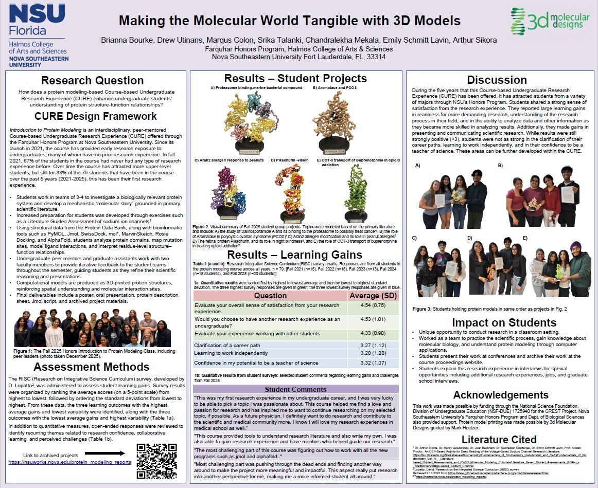
Making the Molecular World Tangible with 3D Models
Authors: Brianna Bourke, Drew Utinans, Marqus Colon, Srika Talanki, Chandralekha Mekala, Emily Schmitt Lavin, Arthur Sikora
Teachers: Emily Schmitt Lavin, Arthur Sikora
School: Nova Southeastern University, Fort Lauderdale, FL
PBD ID: 3FSN