




When Mark Hoelzer and Tim Herman proposed the first colors for our DNA nucleotides, I asked whether the colors mattered. They told me no, so I suggested swapping blue and green so guanine would be green. That swap is in every nucleotide we make today. The colors turned out to matter, because students carry them from one model to the next.
How do consistent colors help students learn from molecular models? When a color means the same thing across kits, students can pick up a new model and already read part of it. That's why we choose colors carefully and why we changed one scheme that no longer made sense.
Molecules have no color at this scale, so every color in a model is a decision someone made. That makes color a good context for practicing evaluating a model's limits, part of NGSS Science and Engineering Practice 2: Developing and Using Models. Color is part of the model, so students should learn to read it.
Where Our Colors Come From
We weigh four factors when choosing a color: the CPK standard, our own history, the colors David Goodsell uses, and the colors already in the kit and related kits.
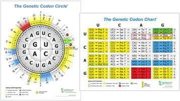
For atoms, we follow CPK: carbon is gray, oxygen red, nitrogen blue, sulfur yellow, and hydrogen white. The Amino Acid Starter Kit© is our gold standard. Each side chain has colored spheres for its atoms and a colored clip for its chemical property, all on a gray backbone. (We explained the property colors in Codon Charts, Color, and Protein Behavior.) From Goodsell's watercolors, we took the background colors of the organelles in the Cell Modeling Kit©.
Nucleotides Keep Their Colors from Kit to Kit
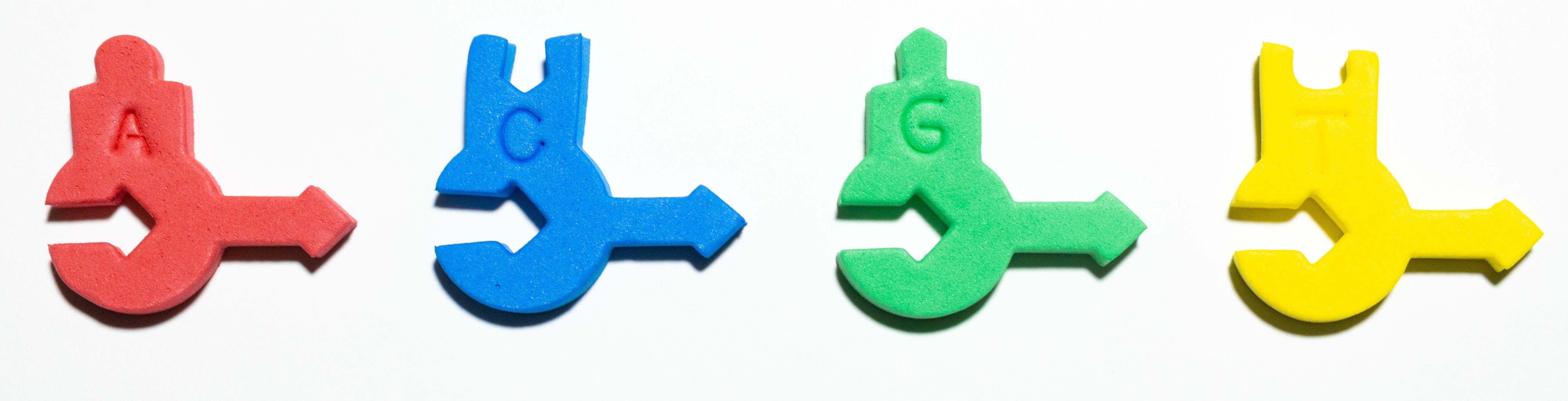
Red adenine, yellow thymine, blue cytosine, and green guanine appear in the DNA Starter Kit©, Flow of Genetic Information Kit©, Nucleotide Student Modeling Pack©, and Biotechnology Kit©. The base labels in the Molecules of Life Modeling Kit© follow the same pattern. Uracil is white because we think of it as a lighter form of yellow, a hint at how closely it is related to thymine.
- Middle school: Students build a short strand with the DNA Starter Kit©. Later, with the Flow of Genetic Information Kit©, they predict the RNA pairing before they see the RNA nucleotides, then explain why uracil might be white.
- High school: In the Biotechnology Kit©, gray nucleotides stand in for unlabeled nucleotides, and the dideoxy nucleotides are gray too. Students explain what the colored and gray pieces each represent in PCR or Sanger sequencing.
Two kits break the pattern on purpose. The Dynamic DNA Kit© uses only CPK colors. The Chromosome Connections Kit© uses its own colors so students can keep the mother's and father's chromosomes separate. Each kit's colors serve its own story.

Membranes Are Always Yellow
For Tim, yellow means hydrophobic, and a membrane's most important feature is its large hydrophobic layer. Every ion and molecule that crosses a membrane has to get through it. So our membranes are yellow in the Phospholipid & Membrane Transport Kit©, Membrane Student Modeling Pack©, Synapse Construction Kit©, Mosaic Membrane Mighty Model© (yellow with red head groups), CRISPR Adaptive Immunity Kit©, and the kits we're field-testing now.
The Phospholipid & Membrane Transport Kit© moves students through three color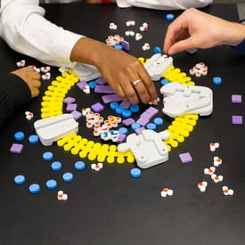 schemes. First, they build a phospholipid in CPK colors. Once they see a hydrophobic region and a mostly negative hydrophilic region, they switch to small yellow phospholipids with red head groups. After they work out how phospholipids arrange into bilayers in water, they get the all-yellow preformed bilayers.
- Middle school: Students place water molecules near a membrane from the Membrane Student Modeling Pack© and explain which part the water stays away from, and why that part is yellow.
- High school: At each switch in the Phospholipid & Membrane Transport Kit© sequence, ask what the new colors keep and what they leave out.
When a Color Scheme Stops Making Sense
For almost 25 years, our antibodies had yellow heavy chains and red light chains. It was one of our first models, and the colors weren't chosen for any particular reason. While developing models for Modeling Infection & Immunity, we realized the scheme no longer matched how we use color, because yellow had come to mean hydrophobic.
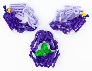 We struggled with it for four years, then dropped it. Antibodies are now a single color that depends on the antigen they block: purple for influenza and red for coronavirus. When a model shows antibody structure, we use two shades of that color, darker for the heavy chain and lighter for the light chain. In the new Dynamic Antibody & Antigen Mighty Model©, that means purple and lavender.
Older antibody models still work. Give students both keys and ask what each scheme helps them see: the antibody's chains or what it binds. A color scheme is a claim about what matters most, and claims can be revised.
Start Each Kit with a Key Check
Across our kits, yellow can mean thymine, a membrane's hydrophobic layer, a hydrophobic side chain, or a sulfur atom. Before students build, ask, "What does yellow mean in this model?"
- Middle school: Set the DNA Starter Kit© next to the Membrane Student Modeling Pack©. Students explain how yellow can mean two different things.
- High school: Students find the methionine side chain in the Amino Acid Starter Kit©, where a yellow sulfur sphere sits on a yellow hydrophobic clip. Which color system does each yellow belong to, and how can they tell?
Your Turn
Try a key check before you open your next kit, and adapt the questions to your students. Which color confuses your students most? Reply and tell us.
Resources for This Lesson
- Amino Acid Starter Kit©
- DNA Starter Kit©
- Flow of Genetic Information Kit©
- Biotechnology Kit©
- Molecules of Life Modeling Kit©
- Phospholipid & Membrane Transport Kit©
- Membrane Student Modeling Pack©
- Mosaic Membrane Mighty Model©
- Cell Modeling Kit©
- Dynamic Antibody & Antigen Mighty Model©
- Codon Charts, Color, and Protein Behavior